Evaluation of Mesothelioma Symptoms
Quick Links
A variety of techniques and technologies may be used to diagnose malignant mesothelioma. The aggressive nature of the disease demands an early diagnosis to maximize treatment options, but its unique behavior pattern and relative rarity among the general population often works against such early identification. In its earliest stages, the cancer typically presents in non-specific ways and is often misdiagnosed because of this non-specific presentation. The majority of patients only receive a diagnosis after the disease has progressed to Stage III or Stage IV. Because of this tragic situation, improvements in our ability to definitively determine a diagnosis are among the most important goals in contemporary research with a large number of studies actively investigating novel diagnostic techniques.
Initial Evaluation of Mesothelioma Symptoms
Pleural mesothelioma is the most common form of the disease, so an initial evaluation of symptoms will likely be due to pulmonary-related issues. It’s important for a physician to learn about any prior asbestos exposures that the patient is aware of so he or she can complete as full a medical history and workup as possible. This type of communication between the patient and doctor is important so that the earliest possible diagnosis can be made: if the doctor does not suspect an asbestos-related disease initially, he or she may not be able to diagnose the disease early enough to maximize prognosis.
In addition to constructing this medical profile and detailed history, a complete physical examination will be performed. During a physical exam your doctor may look for the following:
- Breath Sounds: By listening to the chest cavity as the patient inhales and exhales, the doctor may note differences in the sounds of a patient’s breath from normal breath sounds. This could indicate the presence of a pleural effusion, or another restriction of lung capacity.
- Vocal Sounds: The doctor may listen to the chest cavity while having the patient make vowel sounds – comparing the sound resonance in the right and left chest cavities. Muted, or dull, sounds between the cavities could also be indicative of effusions or another restriction of the lungs.
- By tapping on the chest area a doctor may notice a dull, solid thumping sound, rather than a hollow resonance. This may also indicate the presence of fluid, or a mass, in the chest cavity.
On the basis of patient symptoms, medical history, the presence of specific risk factors—such as work environment and known asbestos exposure—and the results of a complete physical examination, the doctor is likely to schedule one or more of the methods described below to make a mesothelioma diagnosis.
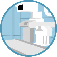
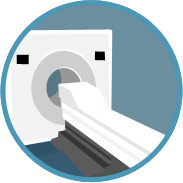
Imaging Technologies
There are many available technologies that allow doctors to view organs and tissue that might show the presence of fluid or tumors. Imaging technologies are important techniques not only to make a mesothelioma diagnosis, but also for determining a treatment plan and for tracking the patient’s response to the treatments.
X-Rays
X-rays are usually the first test given to many patients. X-rays are not as advanced as other tests and their resolution is not as good as some modern tests like the ones described below. Their resolution can only provide “shadows” of where the tumors might be located. X-ray results may help determine if a patient has a pleural effusion or point at the existence of a possible pleural or lung malignancy.
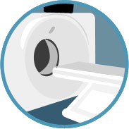
Computed Tomography Scans (CT Scans)
A computed tomography (CT) scan uses x-rays and computers to give more sophisticated and detailed pictures of the insides of the body. CT scans are more advanced that X-ray test and can provide more accurate results.
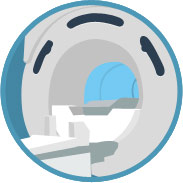
Magnetic Resonance Imaging (MRI)
Magnetic Resonance Imaging or MRI uses magnetic fields and radiowaves to scan the body. MRI provides much better picture than computed tomography scans and is often used for planning patient’s surgery.
Positron Emission Tomography (PET)
Positron Emission Tomography (PET) is an imaging test that help reveal how your tissues and organs are functioning through the use of a radioactive drug (tracer) to show the activity of your organs and tissue and determine if everything is normal or not.

PET-CT (Integrated Positron Emission Tomography – Computed Tomography)
PET-CT is a cutting-edge imaging technique that combines PET scans and CT scans in one machine. These are often called PET scans or PET imaging and is a type of nuclear medicine imaging technology. This test provides the doctor with a more complete understanding of a patient’s individual disease status.
Biopsy Procedures

Laparoscopy
A laparoscopy is a surgical procedure used to obtain a sample of the peritoneal tumor. In this procedure, a flexible tube is attached to a video camera that is inserted into the abdominal cavity via small incisions.

Bronchoscopy
A bronchoscopy is a surgical procedure where doctor uses a viewing tube to evaluate a patient’s lung and airways including the voice box and vocal cord, trachea, and many branches of bronchi. During this process, the surgeon can get small samples of abnormal-appearing tissue may also be removed for testing.

Thoracoscopy
A tissue sample of a pleural or pericardial tumor can be obtained during a thoracoscopy. A thoracoscope, a telescope-like instrument connected to a video camera, is inserted through a small incision into the chest, along with small surgical tools. The doctor can see the tumor through the thoracoscope and can use special tools to take a tissue biopsy.

Mediastinoscopy
As in the bronchoscopy the doctor uses a viewing tube through the chest wall to examine the area between the lungs. This procedure is often recommended to determine how cancerous cells have spread (how far and to which areas) to determine the stage of cancer.